


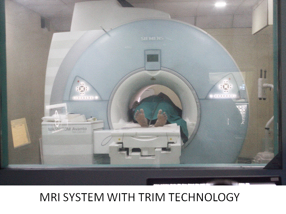
MAGNETOM Avanto 1.5 Tesla 18 Channel MRI System with Tim technology
The MAGNETOM Avanto 1.5 Tesla MRI Scanner with
Tim technology by Siemens Healthcare provides a
new level of seamless image quality with
unprecedented acquisition speed and ease of use.
The heart of the MRI Scanner is the magnet, and
the MAGNETOM Avanto has the best-in-class magnet
homogeneity which results in excellent image
quality. With the MAGNETOM Avanto head-to-toe
imaging in a very short time has come into
routine clinical issue owing to its powerful
hardware and software. The entire system is an
engineering marvel harboring an ergonomically
designed table, gantry and hardware and software
that is designed to ensure the Audio Comfort of
the patient by minimizing the sound levels. This
makes the scan very comfortable for the patient
and helps achieve excellent patient compliance.
It is fully loaded with the neuro, ortho,
cardiac, onco, body, angio, paediatric and
breast suites which ensures uninterrupted
workflow empowering the user to handle all
applications. The new syngo Blade sequence is
extremely useful in minimizing motion induced
artifacts in very sick patients.
Apart from this some advanced applications such
as the following, help the specialists diagnose
critical cases.
syngo SWI – A Siemens unique software which can
diagnose micro-bleeds (diffuse axonal injury)
and hemorrhagic transformation of stroke. The
phase information is useful for detection of
occult vascular disease (cavernomas, angiomas,
and telangiectasias), cerebral venous
thrombosis, intra-arterial clot detection and
other mineral deposition. SWI is a very
significant tool for diagnosing
neurodegenerative diseases (Alzheimer’s,
multiple sclerosis, etc.) and to characterize
tumors.
syngo BOLD: is a comprehensive processing and
visualization package for functional MRI
studies. This allows the visualization of
important brain centers, which control our
activities, and the spatial relationship of
these with normal anatomy or brain lesions. fMRI
studies are helpful in neurosurgical planning,
to assess the effects of neurodegenerative
diseases, trauma or stroke on brain function,
and brain mapping.
syngo Inline Perfusion: It is an important
feature for looking into penumbra in case of
acute stroke which can modify the treatment
planning.
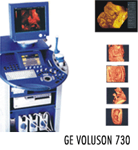
.png) Rajajinagar
Rajajinagar